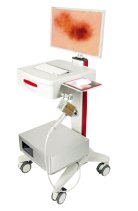
Elegant, robust and flexible – the new system for epiluminescence microscopy and video documentation
dermoscope vexia is the new FotoFinder turn-key system for optimal comfort when working in dermoscopy. The new equipment makes video documentation even more compact, robust and comfortable. Flexible, stable and completely reliable in continuous operation, the device is the perfect complement for practices and clinics alike and brings high technology into patient care. FotoFinder dermoscope vexia is installed ready for use and, thanks to the sophisticated hardware and high-performance SQL database, it is optimally suited to networks.
FotoFinder dermoscope vexia at a glance:
- Turnkey installed turn-key system with FotoFinder quality hardware
- Workstation with stable casters, drawer and mouse pad for left-handed and right-handed operators
- Unrivalled Full HD live quality
- Moleanalyzer with malignancy score (optional)
- Comprehensive reports for your patients
- Fusion of state-of-the-art medical engineering with timeless design
The gold standard for epiluminescence microscopy and video documentation
Full HD live brilliance in screening and follow-up examinations.
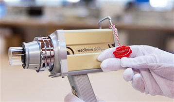
The Full HD video camera medicam 800HD offers incomparable image quality and sets standards in dermoscopy! Robust, reliable and extremely rapid in continuous operation, the camera allows brilliant dermoscopic videos and excellent clinical images, be they of the whole back or individual fingernails. FotoFinder is the only one to have completely integrated the Full HD live image in the software.
The medicam 800HD is a dermoscope component.
- The only system for large panoramic images and dermoscopic images with just one camera
- Maximum video quality in Full HD live with 1,920 x 1,080 pixels
- Unrivalled brilliant Full HD live image integrated in the software
- Patented precision optics with infinitely adjustable optical magnification
- Autofocus at all magnification strengths
- Option of selecting between immersion and polarization
- Flood light for brilliant overview images as "maps" of the nevi and for follow-up documentation
- Spacer for standardized clinical images
- Standardized image parameters
- Attachments for prominent or difficult-to-reach lesions (optional)
- Fluorescence diagnostics attachment (optional)
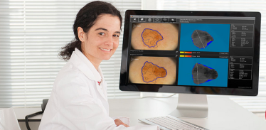
Nevi analysis – tried and tested and reliable!
Computer-assisted second opinion with uniformly high levels of sensitivity and specificity.
In mole analysis, sensitivity and specificity are decisive for how reliable the result is. Sensitivity of far beyond 90% is usually at the expense of specificity and results in increased excision of harmless nevi.
The Moleanalyzer from FotoFinder was developed in cooperation with the University of Tübingen Department of Dermatology and offers equally high sensitivity and specificity. Unlike other systems, it has proven its worth in practice for many years and not just in "trimmed" studies. It is also not intended to replace doctors, but simply to deliver a computer- assisted second opinion based on clinically tested pattern recognition algorithms with a risk score. That offers you – particularly in problematic cases – even more certainty in the early detection of malignant melanomas.
The Moleanalyzer is a Fotofinder dermoscope component.
- Analysis of melanocytic nevi using pattern recognition algorithms
- Reliably high values for sensitivity and specificity
- Impressive visualization of structural changes
- Parallel analysis of two lesions to quantify changes